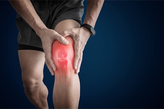
Symptoms OF Arthritis
Patients suffering from arthritis, experience the following symptoms:
- Pain and swelling.
- Joint stiffness with constant ache around the joint.
- Muscle weakness.
- Loss of flexibility.
- Decreased aerobic fitness.
Pathology: There is a loss of collagen and proteoglycans from the matrix. Initially, chondrocytes proliferate and synthesize enhanced amounts of proteoglycan and collagen molecules. As the disease progresses, fibrillation, erosion, and cracking initially appear in the superficial layer of cartilage and progress over time to deeper layers.
Diagnosis Of Arthritis
Physical Examination: The patient is examined is done thoroughly, patient's history is taken. The examination includes observation, palpation, some active movements, and some specific tests.
Thompson Test:Thompson Test is performed during the physical examination. The test is useful for diagnosing complete Arthritiss.
Matles Test: This test is also done during the physical examination. The patient lies in the prone position and is asked to flex the knee up to 90 degrees. Throughout the movement, the examiner observes the feet and ankles. The test is negative when the foot goes slightly into plantar flexion and the test is positive if the foot is in the neutral position or the movement results in dorsiflexion.
Magnetic resonance imaging (MRI): Magnetic resonance imaging (MRI) of the leg is done to diagnose the rupture of the Achilles tendon. MRI scan of the ankle is performed to check for tendon degeneration and assess the severity of the damage.
Achilles Tendon Total Rupture (ATR-score): ATR-score is an important questionnaire that refers to the limitations or difficulties a patient faces who has a tendon rupture.
Real-time Achilles ultrasound Thompson test: This test is like the Thompson test but is done under ultrasound visualization. It can be used by the examiner to provide improved diagnostic characteristics compared with static ultrasound.
X-ray: X-ray of the back of the ankle is taken to rule out other disease conditions.
Ultrasound: The movement of the tendon is observed in real-time. Colour-Doppler ultrasound can also assess blood flow in the region.
Treatment Of Arthritis
Medications: NSAIDs, corticosteroids, analgesics, pain killers, etc.
Note: Medication should not be taken without the doctor’s prescription.
Treatment for a ruptured Achilles tendon depends on the age, activity level, and severity of the injury. Younger and more active people, especially athletes, tend to choose surgery to repair a completely ruptured Achilles tendon, while older people usually opt for nonsurgical treatment.
Surgery: Surgery involves making an incision in the back of the lower leg and stitching the two tendons together. Depending on the severity of the injury of the two tissues, the repair might be reinforced with other tendons. The torn ends of the tendon are reattached with one large incision (open surgery) or many smaller incisions (percutaneous surgery).
Physiotherapy Treatment Of Arthritis
Immobilization: Boots or cast is used for immobilization, the ankle is kept from moving for the first few weeks, usually with a walking boot with heel wedges or a cast, with the foot flexed down.
Cryotherapy: Ice therapy can be used to decrease inflammation and pain.
Ultrasound therapy: Ultrasound therapy is beneficial in the early healing process of tendons when the pulsed mode is used.
Transcutaneous electrical stimulations (TENS): Transcutaneous electrical stimulations (TENS) are found to be effective in the healing of Achilles tendon sutures.
Laser therapy: Laser therapy can be used in the treatment and rehabilitation of Achilles sutures alone or in combination with more conventional therapies.
Range of Motion Exercises: Gentle range of motion exercises like passive ROM to active ROM exercises. Exercises may include Passive ankle ROM in all directions, ankle pumps, ankle active ROM in all directions, ankle alphabet. These motions should be done in a slow and in controlled way for 3 seconds, 15 repetitions up to three times a day.
Flexibility Exercises: Flexibility exercises are done for the ankle and lower extremity, this can help these stretch the tight muscles. The scar tissue that is formed along the injured Achilles can also help remodel the tissue and get it functioning normally, towel calf stretch, stretching for the gastrocsoleus muscles, stair stretching by hanging the heel over the edge of a step, etc. Manual stretching for the muscles surrounding the ankle can also be done. Each stretch should be held for 30 to 60 seconds and performed in 5 to 10 sets.
Progressive Ankle Strengthening: Progressive ankle strengthening exercises are done to improve the strength of the muscles. Placing progressive stress on the Achilles tendon with strengthening exercises, helps to improve the overall function. Manual resistance, calf raises, short arc quad sets, and straight leg raise, these exercises can be done 10 to 15 times, in 2 -3.sets.
Balance and Proprioception: Balance and proprioception impairments may develop after Arthritis, due to the period of immobilization. Therefore specific exercises are recommended to improve the balance. E.g. single-leg stance, wobble board exercises, standing on foam, etc.
Plyometrics: Plyometrics or high-level sports and recreational activities are also a part of the rehab program. Plyometrics are specific exercises that require to quickly jump and land in a specific and safe manner, e.g. are hopping in place, over lines, forward and backward, and side to side, first with two feet, and then with the affected foot only, etc.
Aerobic Conditioning: The physiotherapist recommends aerobic conditioning to counteract the deleterious effects of immobilization. Aerobic exercises help to improve the strength, from non-weight bearing with minimal or no impact to full weight bearing with some impact. These exercises include biking, elliptical trainer, treadmill walking, and running.
To get the maximal effect, these exercises should be done for 30 minutes for 5 days a week. These exercises aim to return to the highest level of performance, as required by an athlete or during everyday life.
Patient Education
The patient is advised to stop the exercises that cause pain or swelling around the Achilles tendon. This may be a sign of overdoing it and may need to rest a bit before continuing. And is also advised to continue strength and stability exercises even after the recovery because some problems can persist for up to a year.