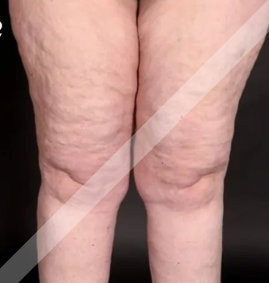
Call Whatsapp

Lipedema

What is Lipedema?
Lipedema is a chronic, progressive disorder of fat metabolism that primarily affects women and is characterized by a symmetrical and disproportionate accumulation of fat, typically in the lower limbs (hips, thighs, buttocks, and legs) and sometimes in the arms. This fat distribution usually spares the hands and feet and is often associated with pain, tenderness, easy bruising, and swelling that does not respond to diet or exercise. The condition is believed to have a hormonal and genetic component and is often misdiagnosed as simple obesity or lymphedema.
It may worsen over time and can significantly impact mobility and quality of life if not properly managed.
Symptoms OF Lipedema
Mentioned below are the common symptoms of Lipedema:
  1. Disproportionate fat accumulation: Excess fat mainly in the hips, thighs, buttocks, and legs (sometimes arms), with the feet and hands typically spared.
  2. Symmetrical fat distribution: Both sides of the body are affected equally.
  3. Painful fat: The affected areas are often painful or tender to touch, even without pressure.
  4. Easy bruising: The skin bruises easily due to fragile blood vessels in the fat tissue.
  5. Swelling (edema): Legs or arms may swell, especially as the day progresses, often worsening with prolonged standing or sitting.
  6. Skin changes: Skin over the affected areas may feel soft, cool, and have a dimpled or nodular ("orange peel") texture.
  7. Fat that is resistant to diet and exercise: The fat deposits do not reduce significantly with weight loss efforts.
  8. Restricted mobility: Heaviness and discomfort can lead to difficulty in walking or standing for long periods.
  9. Emotional distress: Body image concerns and chronic discomfort often lead to depression, anxiety, or social withdrawal.
  10. Progressive worsening: Without treatment, the condition may progress over time, sometimes leading to secondary lymphedema (known as lipo-lymphedema).
Causes of Lipedema
The exact cause of lipedema is not fully understood, but several contributing factors have been identified. These include:
  1. Hormonal Factors: Lipedema almost exclusively affects women and often begins or worsens during times of hormonal change such as:
    1. Puberty
    2. Pregnancy
    3. Menopause
    This suggests a strong hormonal influence, particularly involving estrogen.
  2. Genetic Predisposition: Lipedema often runs in families, indicating a hereditary component. Up to 60% of patients report a family history of similar symptoms.
  3. Microvascular Dysfunction: Dysfunction in small blood vessels and capillaries may contribute to increased leakage of fluid and fragile blood vessels, leading to fat accumulation and bruising.
  4. Connective Tissue Disorders: Some researchers suggest that abnormalities in connective tissue may play a role, affecting the structure and function of the fat and lymphatic systems.
  5. Lymphatic System Involvement (Secondary): Although lipedema is distinct from lymphedema, in later stages, impaired lymphatic drainage may develop (called lipo-lymphedema), worsening swelling and symptoms.
  6. Inflammation: Chronic low-grade inflammation may be present in lipedematous tissue, contributing to pain and progression of the disease.
Diagnosis Of Lipedema
Here is a list of commonly used diagnostic techniques for lipedema:
  1. Clinical Examination:
    This is the primary method of diagnosing lipedema.
    Visual inspection and palpation of fat distribution (especially legs, hips, buttocks, arms).
    Noting symmetry, tenderness, and easy bruising.
    Assessing if hands and feet are spared (important to differentiate from lymphedema).
    Checking for non-pitting edema and pain on pressur
  2. Detailed Medical History:
    Onset during puberty, pregnancy, or menopause.
    Family history of similar symptoms.
    History of diet-resistant fat gain in specific areas.
    Symptoms such as pain, swelling, and easy bruising.
  3. Stemmer’s Sign Test:
    A simple test to differentiate lipedema from lymphedema.
    If you can pinch and lift a fold of skin at the base of the second toe/finger, Stemmer’s sign is negative, which supports lipedema.
    A positive Stemmer's sign (inability to pinch skin) suggests lymphedema.
  4. Imaging Techniques: Used to rule out other conditions or support the diagnosis.
    1. Ultrasound (Sonography):
      Can show abnormal fat structure and fluid retention.
      Helps rule out venous insufficiency.
    2. MRI or CT Scan:
      Provides a detailed view of fat and soft tissue distribution.
      Can differentiate normal vs. pathological fat.
    3. Lymphoscintigraphy:
      A nuclear imaging test used if lymphatic involvement is suspected.
      Often normal in early lipedema, but can show changes if lipo-lymphedema has developed.
  5. Body Composition Analysis:
    Measures fat distribution and volume.
    Can support diagnosis, especially when combined with clinical findings.
Treatment Of Lipedema
Medications: Ibuprofen, naproxen, paracetamol, pregabalin, gabapentin, etc
Note: Medication should not be taken without the doctor’s prescription.
Physiotherapy Treatment Of Lipedema
Physiotherapy plays a key role in the conservative management of lipedema. While it cannot eliminate the abnormal fat deposits, it helps manage pain, swelling, reduced mobility, and fatigue, and improves functional ability and quality of life.
  1. TENS: TENS is used to reduce pain, by stimulating the release of endorphins.
  2. Interferential Therapy (IFC): IFC stimulates deeper tissues, reducing pain and inflammation.
  3. Therapeutic Ultrasound: Therapeutic Ultrasound helps to generate heat and promote tissue healing.
  4. Manual Lymphatic Drainage (MLD):
    1. A gentle, rhythmic massage to stimulate the lymphatic system
    2. Reduces fluid retention, pain, and tightness
    3. Usually done 2–5 sessions/week depending on stage
    4. Can be taught to the patient for self-MLD
  5. Compression Therapy:
    Use of medical-grade compression garments (stockings, sleeves)
    Prevents fluid buildup, supports tissues, reduces discomfort
  6. Exercise Therapy:
    1. Low-Impact Cardiovascular Exercise
      To stimulate lymphatic flow and improve circulation:
      1. Aquatic therapy (swimming, walking in water)
      2. Cycling (stationary or regular)
      3. Brisk walking
      4. Rebounding (mini trampoline) – if tolerated
    2. Resistance Training
      1. Builds muscle tone and improves function
      2. Focus on lower limbs and core
      3. Use light weights or resistance bands
    3. Stretching and Mobility
      1. Gentle stretching of calves, hamstrings, and hips
      2. Improves flexibility and reduces stiffness
  7. Lymphatic Taping (Kinesio Taping)
    1. Special taping technique used to:
    2. Support lymph drainage
    3. Reduce pain and heaviness
    4. Improve movement awareness
  8. Education & Self-Management Training
    1. Posture correction and body mechanics
    2. Importance of regular movement, especially in sedentary jobs
    3. Skin care to prevent infections (like cellulitis)
    4. Teaching self-MLD and home exercises
    5. Encouraging consistent use of compression garments
  9. Psychological and Behavioral Support
    1. Physiotherapists can help identify:
    2. Anxiety, low mood, or body image distress
    3. Refer to psychological or support services
    4. Encourage group exercise or support group participation