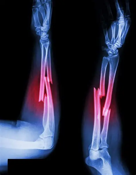
What is Forearm Fractures?
Forearm fracture is the fracture, which occurs between the wrist and the elbow. The bones which get fractured are the radius and the ulna. Radius bone lies on the thumb side of the forearm whereas the ulna bone lies on the little finger side.
Fractures can occur in one or both bones at one or several places of the forearm:
- Middle of the Forearm
- Near the wrist, the distal end of the bone.
- Near the elbow, the proximal end of the bone.
Types of Forearm FracturesForearm fractures can occur in the radius or ulna or in both bones.
- Torus fracture: This type of fracture is a stable fracture, the broken bone pieces are still in position and not displaced.
- Metaphyseal fracture: The fracture occurs across the upper or lower portion of the bone without affecting the growth plate.
- Greenstick fracture: The fracture extends through the length of the bone and causes it to bend on the other side.
- Galeazzi fracture: This fracture affects both bones of the forearm. Such fractures are usually displaced fractures in radius and dislocation of the ulna at the wrist.
- Monteggia fracture: This fracture affects both bones of the forearm. These fractures usually occur in the ulna and the head of the radius is dislocated.
- Growth plate fracture: This fracture occurs at the growth plate of the radius near the wrist.
Symptoms OF Forearm Fractures
A forearm fracture usually results in:
- Severe Pain
- Swelling
- Discolored skin around the affected area
- Feeling of numbness.
- Loss of forearm and hand function
Pathology: Fracture depends upon the age of the individual, the strength of the bone, mass of the bone, quality of the bone, and the frequency, nature, and forces produced by the injury on the bone. Usually repetitive or high-velocity thrust leads to mechanical failure.
Diagnosis Of Forearm Fractures
X-rays: X-rays show clear images of the bones and help to determine the extent of the injury.
MRI: Magnetic resonance imaging (MRI) creates very detailed images using magnetic fields.
CT scan: Computed tomography scan (CT scan) provides detailed cross-sections of the bone.
Bone scan: A bone scan helps to find fractures that are not shown up in an X-ray, it can help find those fractures.
Treatment Of Forearm Fractures
Depending on the type of fracture and degree of displacement, treatment is decided.
Nonsurgical Treatment: Undisplaced fractures may simply need the support of a cast or splint while they heal.
Severe fractures that have become angled, gentle push or manipulation (closed reduction) of the bones into place is done.
span>Surgical Treatment: Surgery is required in displaced fractures or maligned fractures to align the pieces of bones and secure them in place. In such fractures, the skin is opened and the broken bone segments are repositioned (open reduction). Metal implants, pins, stainless-steel screws, plates, and fixators, or a cast is used to hold the broken bones in place.
Conservative Treatment For Forearm Fractures
Medications: Pain reliever, such as acetaminophen, ibuprofen etc
Note: Medication should be taken under the prescription of the doctor.
Recovery: A stable fracture may require 3 to 4 weeks in a cast. Whereas severe fractures may require immobilization for 6 to 10 weeks.
After the removal of the cast, the wrist and elbow joints may be stiff for 2 to 3 weeks. Physiotherapy is recommended after the cast removal.
Cryotherapy: Cryotherapy is used to help reduce pain and swelling.
TENS: Electrical stimulation (TENS) may also be used to help improve muscle and strength.
Ultrasound: Ultrasound helps to reduce the scar adhesion thus improving mobility.
Massage: After a surgical intervention scar massage and mobilization can help reduce adhesions. Massage helps to improve mobility around the scar.
Range of motion exercises: Range of motion exercises help to regain full motion of the elbow, wrist and rotation of the forearm. Early range of motion exercises are started from distal to proximal. Particular focus should be placed on the fractured area and the joints around it. Exercises like simple flexion, extension, supination, pronation exercises can be done with the help of wall pulleys, shoulder wheels, finger ladder etc.
Proprioception: Proprioception is the ability to know where the body is, with eyes closed. Due to immobility proprioception decreases. To enhance the sense of proprioception exercises like pushups, rolling a ball on a surface with hand, holding up weight overhead while moving the shoulder all the exercises can be done as a training.
Strengthening exercises: Initially, physiotherapy may help to improve the strength of the unaffected limbs, to prevent the stiffness in joints around the fractured area. Shoulder wheel with resistance, weight cuffs, dumbells, resistance bands, resistance tubes, and other types of equipment may be used to improve strength and mobility of the muscles and joints and around the fractured area. Initially Exercises are done to retrain the joint and muscles that the bone can tolerate the loads and stresses while doing everyday functions. Physiotherapy can help to return to optimum function depending on the severity of the injury.
High-intensity exercises: These are advanced exercises such as doing quick movements, throwing a ball, catching, lifting, etc., and progressively increased resistance exercises are done to increase the overall functional mobility.